A 16-year-old boy with fever (38.9°C) and sore throat for the last 8 days was unresponsive to antibiotics. He was febrile and anemic and had generalized lymphadenopathy and hepatomegaly. Oral examination revealed painful, bilateral extraoral herpetiform lesions adjacent to the vermilion border of the lower lip (Fig. 1). These lesions appeared as clusters of encrusted vesicles. Intraoral ulcerations, suggestive of herpes simplex infection were present on the marginal and interdental gingiva on the buccal and palatal aspects of all teeth as well as on the lateral borders of the tongue (Figs. 2 and 3). These deep, “punched-out” ulcerations were covered with curdy, milky-white patches suggestive of oral candidiasis. The colour of the gingiva varied from pale to purplish red; loss of normal contour and stippling of gingiva and purple bruising were observed on the posterolateral aspect of the palate (Fig. 2). Candidiasis was also noticed on the posterior third of the tongue (Fig. 3). The pericoronal gingivae on the erupting mandibular left and right second molars were inflamed and covered with oral thrush (Fig. 4). There were generalized stains and calculus.
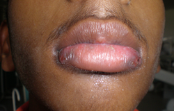 Figure 1: Bilateral extraoral herpetiform lesions with vesicles and encrustation.
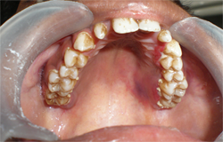 Figure 2: Palatal bruising; markedly swollen, friable and erythematous gingiva; soft, raised, curdy, milky-white plaques interdentally suggestive of candidiasis.
 Figure 3: Ulceration, candidiasis and bruising (ecchymosis due to hemorrhage) on the tongue.
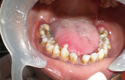 Figure 4: Herpetic ulcers; enlarged, edematous, reddened pericoronal flaps superimposed with candidiasis, which is evident on the tongue and interdental areas.
What is the condition?
Differential Diagnosis
The differential diagnosis included immunodeficiency, hematologic malignancies, viral infection, immunosuppressive therapy, genetic causes and systemic conditions, including aplastic anemia, systemic lupus erythematosus and cyclic neutropenia.
States of Immunodeficiency
Defects in the functioning of the immune system are characterized by increased susceptibility to frequent, severe and recurrent infections of the respiratory tract, skin and mucous membrane. These are classified by type of cell affected, i.e., defects of B-lymphocytes, T-lymphocytes, phagocytic cells or a complement cascade. Infections with major gram-positive organisms occur frequently in B-cell immunodeficiencies, whereas severe viral and fungal infections occur in T-cell immunodeficiencies.
Acquired immunodeficiencies are the result of extrinsic causes, such as malnutrition, aging and medications, e.g., cancer chemotherapy, steroids and immunosuppressive drugs used by organ transplant recipients and in auto-immune disease. Chronic infections, such as AIDS caused by HIV, also impair the functioning of the immune system. HIV destroys CD4 helper/inducer T cells and affects both humoral and cell-mediated immunity making the patient highly susceptible to opportunistic infections. Manifestations of overall progression of the disease are more common in children because of their immature immune system. A prolonged history of fever, chronic recurrent diarrhea, oral hairy leukoplakia and Kaposi’s sarcoma may be some of the differentiating factors. CD4+ T-lymphocyte values should be obtained if a child has a positive virologic test for HIV.
Hematologic Malignancies
This group of immunocompromised states may result from an underlying hematologic malignancy or the use of chemotherapeutic drugs. Myeloid leukemias are characterized by infiltration of blood, bone marrow and other tissues by neoplastic cells of granulocytic series; in lymphoblastic leukemias, the bone marrow is replaced with small, immature lymphoblasts. The diagnosis is established by bone marrow aspiration and cytology, immunophenotyping, immunohistochemistry, molecular analysis and cytogenetic analysis. Lymphomas are usually diagnosed after microscopic examination of lymph node biopsies, immunohistochemistry and the absence of peripheral blood involvement.
Viral Infection
Infectious mononucleosis, caused by Epstein-Barr virus, is characterized by fever, lymphadenopathy, hepatosplenomegaly and abnormal blood lymphocytes. It can be differentiated from acute leukemia by the distinctive morphology of reactive lymphocytes and lymphoblasts and elevated viral titers. Moreover, in viral disease, lymph nodes, liver and spleen are often soft and ill-defined, whereas in acute leukemia, they are firm and discrete.
Immunosuppressive Therapy
Immunosuppressive drugs include glucocorticoids and cancer chemotherapeutic agents; these produce deficiencies in the host defense system and make a child more prone to infections by opportunistic organisms.
Genetic Causes
Wiskott-Aldrich syndrome (WAS) is an inherited disorder of the immune system that affects only males and is characterized by recurring severe opportunistic infections, eczema and thrombocytopenia. The diagnosis is confirmed by demonstrating a decrease or absence of the WAS protein in blood cells or the presence of a mutation in the gene that codes the WAS protein. Moreover, WAS platelets are significantly smaller than normal platelets.
Systemic Conditions
Aplastic anemia is a rare blood dyscrasia, in which peripheral blood pancytopenia results from reduced or absent blood-cell production in the bone marrow, and normal hematopoietic tissue in the bone marrow is replaced by fatty marrow. Sepsis and hemorrhage are the main causes of death in these patients as a result of neutropenia and thrombocytopenia, respectively. The lymphadenopathy, hepatosplenomegaly and skeletal changes associated with leukemia are not seen. Bone marrow aspiration or biopsy confirms the diagnosis.
Systemic lupus erythematosus is a multi-organ system autoimmune disease characterized by widespread vasculitis and the presence of various auto-antibodies. Fever, joint symptoms, mild anemia and oral ulcers are usually present in active disease. Children with lupus frequently have lymphadenopathy and hepatosplenomegaly. Non-erosive, symmetric and polyarticular arthritis, typical erythematous “butterfly” rash over the malar area and discoid rash with a history of photosensitivity over the face and chest are pathognomonic. Prominent laboratory findings include lymphopenia, thrombocytopenia, low serum complement levels, a high-titered positive antinuclear antibody test and abnormal urinary sediment.
Cyclic neutropenia, an autoimmune disease, is characterized by cyclic fluctuations in neutrophil count. The cycle averages 21 days (14–36 days) with severe neutropenia (neutrophil count < 0.2 × 10⁹/L) lasting 3–10 days. During the period of neutropenia, bone marrow aspirate shows signs of maturation arrest at the myelocytic stage. Cyclic neutropenia is associated with fever, recurrent oral ulcerations, pharyngitis, cervical lymphadenopathy and skin lesions. Intraoral features include severe recurrent gingivitis, loss of periodontal attachment, deep periodontal pocket formation and marked alveolar bone loss, often causing loss of teeth.
Diagnosis of Patient
The patient’s history and clinical examination suggested a hematologic malignancy, and this diagnosis was further corroborated by results of a peripheral blood smear. The hemogram revealed hemoglobin level of 87 g/L, platelet count of 48 × 109/L, elevated total leukocytic count (210.5 × 10⁹/L) and an increase in the number of myeloblasts (98% of the total leukocyte count) with a concomitant decrease in the lymphocyte count (2% of the leukocyte count). The peripheral blood smear revealed severe neutropenia (absolute neutrophil count [ANC], 0 × 109/L). Bone marrow biopsy and immunophenotyping confirmed the diagnosis of minimally differentiated acute myeloid leukemia.
Early bone marrow transplantation is the treatment of choice for acute myeloid leukemias. The patient has been undergoing chemotherapy at a nearby hematology centre and is currently in the consolidation phase. He is awaiting bone marrow transplant.
Leukemia is accompanied by febrile neutropenia, which is defined as the presence of fever (≥ 38.3°C) and a low neutrophil count. Neutropenia is classified as mild (ANC 1.0–1.5 × 10⁹/L), moderate (ANC 0.5–1.0 × 10⁹/L) or severe (ANC < 0.5 × 10⁹/L). Severe neutropenia increases susceptibility to bacterial or fungal infections and impairs resolution of these infections. Leukemia also brings about defects in cell-mediated immunity that may increase the possibility of infection by an inherently present latent virus, such as herpes simplex virus (HSV), which manifests on the oral and circumoral mucosal surfaces. Confirmation of both herpetic gingivostomatitis and candidiasis by appropriate laboratory means is mandatory, even when prior empirical treatment is being contemplated.
Systemic antifungal therapy is indicated for patients who are at high risk of candidemia or are relatively resistant to topical antifungal agents. Nystatin in the form of a mouthwash or suspension or clotrimazole troche can be recommended for the treatment of oral candidiasis. If oral candidiasis is accompanied by an immunocompromised state, fluconazole or caspofungin is effective. The prophylactic use of the antiviral drug acyclovir is known to reduce the incidence of oral infections caused by HSV in patients with acute myeloid leukemia. Patients afflicted with hematologic malignancies are at a high risk of developing life-threatening septicemia due to odontogenic bacteria. Dental extractions and any other oral surgical procedures should be performed at least 3 weeks before the start of cancer chemotherapy. An ANC greater than 1.0 × 10⁹/L and a platelet count of at least 60 × 109/L are acceptable for performing oral surgery.
This case demonstrates the importance for dental practitioners of recognizing mucocutaneous manifestations of systemic diseases. The oral cavity is an important portal of entry for microorganisms, which, in immunosuppressed patients, can lead to a systemic involvement accompanied by life-threatening complications. If such lesions are undetected and untreated, they often lead to a fulminant infection causing death. Dental prophylaxis and treatment of caries and gingivitis are necessary to eliminate the oral source of infection.
THE AUTHORS
|
Dr. Sharma is assistant professor, department of pedodontics, HSJ Institute of Dental Sciences and Hospital, Panjab University, Chandigarh, India. |
|
|
Dr. Bhalla is assistant professor, department of pedodontics, HSJ Institute of Dental Sciences and Hospital,Panjab University, Chandigarh, India. |
Correspondence to: Dr. Urvashi Sharma, House Number 433, Sector 37-A, Chandigarh (India), 160036. Email: navneet207@rediffmail.com
The authors have no declared financial interests.
This article has been peer reviewed.
References
- Whitlock JA, Gaynon PS. Acute lymphoblastic leukemia in children. In: Greer JP, Foerster J, Lukens JN, editors. Wintrobe’s clinical hematology. 11th ed. Lippincott: Lippincott Williams & Wilkins; 2003. p. 1752-74.
- Dupuis-Girod S, Medioni J, Haddad E, Quartier P, Cavazzana-Calvo M, Le Deist F, et al. Autoimmunity in Wiskott-Aldrich syndrome: risk factors, clinical features, and outcome in a single-center cohort of 55 patients. Pediatrics. 2003;111(5 Pt 1):e622-7.
- Sepúlveda E, Brethauer U, Rojas J, Le Fort P. Oral manifestations of aplastic anaemia in children. J Am Dent Assoc. 2006;137(4):474-8.
- Stichweh D, Pascual V. [Systemic lupus erythematosus in children]. An Pediatr (Barc). 2005;63(4):321-9. [Article in Spanish]. Erratum in: An Pediatr (Barc). 2005;63(5):468.
- Hughes WT, Armstrong D, Bodey GP, Bow EJ, Brown AE, Calandra T, et al. 2002 guidelines for the use of antimicrobial agents in neutropenic patients with cancer. Clin Infect Dis. 2002;34(6):730-51. Epub 2002 Feb 13.