A 20-year-old male patient presented with swelling and pain in the right maxillary lateral incisor region. Intraoral examination revealed that the permanent upper lateral incisors were unerupted and the deciduous upper lateral incisors were still present. The crown of the right lateral incisor was dark compared with adjacent teeth, cone-shaped and showed a fissure and caries in the central region (Fig. 1).
Radiologic evaluation confirmed that the permanent upper lateral incisors were missing and the deciduous right upper lateral incisor had unusual radiographic features—a structure expanding toward the apex and surrounded by periapical radiolucency (Fig. 2).
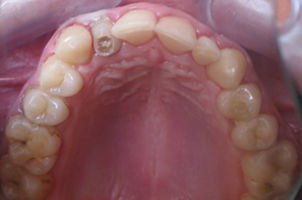 Figure 1: The deciduous right upper lateral incisor, showing dark colouration and caries.
 Figure 2: Periapical radiographic view of the deciduous upper lateral incisor, showing a structure expanding toward the apex and surrounded by periapical radiolucency.
What is this condition?
Discussion
The maxillary right lateral incisor was diagnosed as type III dens invaginatus according to Oehlers' classification.1
After consultations with the department of endodontics and the department of prosthodontics, it was decided that the tooth could not be restored and the patient would receive a dental implant following extraction. To avoid excessive bone loss, the tooth was first separated, then removed. After extraction of the tooth, the socket was thoroughly curetted.
Dens invaginatus is a rare malformation of teeth, probably resulting from infolding of the dental papilla during tooth development.2 The frequency of the condition varies from 0.04% to 10%.3 Oehlers1 categorizes invaginations into 3 classes, based on how far the invaginations extend radiographically from the crown into the root.
Type I is an enamel-lined minor invagination not extending beyond the amelocemental junction. Type II consists of an enamel-lined form that invades the root, ending as a blind sac. Type III involves extension of the enamel-lined invagination through the root to form an additional apical or lateral foramen.
Although the literature includes a few reports of dens invaginatus involving primary teeth,4,5 we did not find cany case of a type III dens invaginatus in deciduous dentition.
In the past, various techniques have been proposed to manage type III dens invaginatus, including conservative restorative treatment, nonsurgical root canal treatment, endodontic surgery, intentional replantation and extraction.6 Considering the anatomy of the tooth and the abnormal dentition process, we decided to remove the tooth surgically.
We believe that, if possible, dens invaginatus in deciduous dentition should be examined carefully and treated promptly as delayed intervention may cause complications, such as periapical infections or patient disturbance.
THE AUTHORS
|
Dr. Okcu is an assistant professor in the department of oral and maxillofacial surgery, Gülhane Military Medical Academy, Ankara, Turkey. |
|
|
Dr. Erdemci is a PhD student in the department of oral and maxillofacial surgery, Gülhane Military Medical Academy, Ankara, Turkey. |
|
|
Dr. Gulses is a PhD student in the department of oral and maxillofacial surgery, Gülhane Military Medical Academy, Ankara, Turkey. |
|
|
Dr. Sencimen is an assistant professor in the department of oral and maxillofacial surgery, Gülhane Military Medical Academy, Ankara, Turkey. |
Acknowledgements: The authors would like to thank Dr. Gunhan, a professor in the department of pathology, Gülhane Military Medical Academy, for his help with this manuscript.
Correspondence to: Dr. Aydin Gulses, Gülhane Military Medical Academy, Department of oral and maxillofacial surgery, General Tevfik Sağlam Cad. 06018, Etlik , Ankara, Turkey. Email: aydingulses@gmail.com
The authors have no declared financial interests.
This article has been peer reviewed.
References
- Oehlers FA. Dens invaginatus (dilated composite odontome). I. Variations of the invagination process and associated anterior crown forms. Oral Surg Oral Med Oral Pathol. 1957;10(11):1204-18.
- Hulsmann M. Dens invaginatus: aetiology, classification, prevalence, diagnosis and treatment considerations. Int Endod J. 1997;30(2):79-90.
- Jung M. Endodontic treatment of dens invaginatus type III with three root canals and open apical foramen. Int Endod J. 2004;37(3):205-13.
- Rabinowitch BZ. Dens in dente: primary tooth; report of a case. Oral Surg Oral Med Oral Pathol. 1952;5(12):1312-4.
- Holan G. Dens invaginatus in a primary canine: a case report. Int J Paediatr Dent. 1998;8(1):61-4.
- Kusgoz A, Yildirim T, Kayipmaz S, Saricaoglu S. Nonsurgical endodontic treatment of Type III dens invaginatus in maxillary canine: an 18-month follow-up. Oral Surg Oral Med Oral Pathol Oral Radiol Endod. 2009;107(3):e103-6. Epub 2009 Jan 25.