Background
Today, a large proportion of practitioners, including ever-increasing numbers of dental students, avail themselves of surgical magnification in daily practice, to improve both their vision and their posture. Higher magnification entails a need for increased lighting at the clinical site.1
Headlamps were used by dentists as early as the 1950s. Since the 1990s, small head-mounted lighting devices with vastly improved mechanical and electronic features have become available to dentists,2 and during the past decade, LED (light-emitting diode) technology has been brought to bear. In addition to decreasing the weight and heat associated with earlier models of head-mounted lights, high-intensity LED lights (Fig. 1) have also greatly increased the portability and battery life of surgical lighting units, especially through the use of lightweight lithium ion batteries.
A head-mounted or spectacles-mounted LED light source has several advantages over other types of lighting sources for the oral working field. These advantages include mobility for free movement from one operatory to another (i.e., the clinician is no longer attached by a cord to an electrical outlet or fibreoptic light source), greater intraoral shadow-free visibility, and moderate cost of purchase and maintenance. LED lights operate at a low temperature, and bulb replacement is unlikely to be required.3 One recent study suggested that white LEDs are better than halogen light sources for evaluation of tooth colour.4
Critical Features for Head-Mounted LED Lights
So what should you look for in a head-mounted or spectacles-mounted LED light? There are several critical features to consider, regardless of whether you are planning to mount the light on your loupes (surgical telescopes), on a headband, or on a face shield. Of course you will need to ensure that the light can be attached and adjusted conveniently, so that it can be focused on your optimal operating control point for intraoral care. That point will likely be located in your midsagittal plane, at about heart height. You should not acquire a light that could force you to compromise your optimal working position.5,6
The adjustability of light intensity is important. Is brighter better? No. You must be able to adjust the intensity of the light to allow for variations in background lighting and level of magnification. Glare and the target-to-background brightness ratio must be controllable to minimize eye fatigue and optimize the ability to make visual discriminations. A range of 10 000 to 15 000 lx (about 900 to 1400 foot-candles) is referenced as an adequate range of light on field if the target-to-background brightness ratio is properly maintained. This level is far exceeded by most LED lights, which is why adjustability is so important.7
To assess the properties of the light and light field generated by the unit, set a plain piece of white paper at your optimal working distance and observe these properties (as described below) on the paper, both with the naked eye and with magnified viewing. It is also useful to observe the field with background lights at different intensities, first turned down and then turned up more brightly. Similarly, it is worthwhile to observe the light generated through the entire range of intensity allowed by the unit you are evaluating.
What colour is the beam? Most manufacturers claim that the newer LEDs are “bright white.” You will have to be the judge as you examine each individual light in your own operatory settings.
The “ideal” size of the lighted field depends on the magnification that will typically be used. The lighted field should be just as large as the magnified field and no more, to diminish the risk of inadvertent flash or glare in the patient’s or the assistant’s eyes.
Look for a distinct field edge. Poor field-edge definition means that the edges of the lit field are vague, offering blends of colour or intensity, or there may be a band of partial light between the lit and unlit areas (Fig. 2). Like a field size that is just large enough, crisp field edges are important to minimize the risk of glare in the patient’s eyes, as well as the risk of flash reflections into the assistant’s eyes. The character of the field edge can be easily seen when the light is focused on a plain sheet of white paper at the optimal working distance.
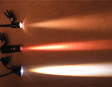 Figure 1: Photograph showing the light beams from 3 different light units illustrates clearly that not all surgical head-mounted lights are created equal.
 Figure 2: An example of a light field with unacceptably poor edge characteristics.
Is there true edge-to-edge clarity of the field? Look for complete homogeneity of the lit field. Unevenness of the field characteristics means that the lit area is not homogenous in colour and/or texture. For example, note the particularly uneven central areas of the 2 circular light fields in Fig. 3. The fields of many lights have wavy or straight lines or a pebbled texture. These characteristics can detract from the clarity of visual perception. Such properties are not always obvious if the unit is tested with a dark or varied background, a situation in which the clinician’s assessment of this important quality will be more difficult.
How easily can the light be cleaned and disinfected? In general, LED units are better sealed and are more compact to allow easier cleaning than was the case with previous light types, but the same cautions apply as for all personal equipment. To comply with typical current asepsis protocols, a bag-type or sticky-strip barrier on the unit housing the battery and switch (or overgloves) may be required to adjust the light intensity if such adjustment is required during the course of a treatment (Fig. 4).
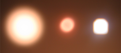 Figure 3: Characteristics of the light field. Note the variation in colour, pattern and size of field for each of the 3 lights.
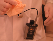 Figure 4: An overbarrier may be required for adjusting light intensity during a procedure.
What about the use of LED headlights when placing light-cured restorative materials? If you work with composite resins, be aware that exposure to any type of light can cause premature curing. To avoid this problem, you can turn the light down or off, or you can use optional amber filters, which are available from some manufacturers.
What is the expected charge life of the battery? These days, a period of use as long as 8–12 hours per charge (or more) is common, but there are still several important questions to ask about the battery. Is the charge life computed for usage of the light at its highest or lowest intensity? What is the degradation curve for charge retention if the LED is in frequent use? Does the battery require full discharge before it can be recharged to full capacity? What is the cost for replacement or additional batteries? Who sells them?
Other issues that you might want to consider are more likely to be matters of personal choice, and many brands offer reasonable solutions and options:
- weight and comfort of the battery pack, especially the intended mode of carrying (e.g., belt, pocket, shirtsleeve)
- convenience, in terms of length and deployment of the wire connecting the battery pack to the light
- convenience of access to the on–off and intensity switch (or switches)
- directional adjustability (little should be required if the mounting is stable and the original set-up accurate)
- availability of adapters for your brand of loupes (surgical telescopes), either from the manufacturer of the LED light or from the manufacturer of your loupes
Head-mounted LED lights for use in a dental practice are available with a wide range of features and in a broad price range ($200 to $1500), and most designs are being upgraded, revised and replaced frequently as LED technology becomes more inexpensively accessible. But, as is the case for so many other instruments and devices, some designs compromise excellence in the delivery of technology most suitable for specific surgical applications. New LED lights become available every week, and many dental manufacturers and suppliers have been phasing out lines every few months as newer, cheaper technology comes available. However, the replacements have not been uniformly better products, according to the critical features described above.
A final piece of advice: Do not buy LED lights online, particularly from unknown industry sources, unless you are prepared to do some return-shipping if the particular light sent to you does not meet one or more of the critical criteria. In some ways, LED technology is still somewhat primitive when used for surgical applications. Furthermore, for many currently available products, quality control is abysmal. Units with uneven fields of illumination can be a particular problem even among identically labelled and manufactured lights from the same company. Buyer beware!
THE AUTHOR
|
Dr. Rucker is professor and chair of general dentistry in the department of oral health sciences, faculty of dentistry, University of British Columbia, Vancouver, British Columbia. |
Correspondence to: Dr. Lance Rucker, Faculty of dentistry, Nobel Biocare Oral Health Centre, 2151 Wesbrook Mall, Vancouver, BC V6T 1Z3. Email: lrucker@interchange.ubc.ca.
The author has no declared financial interests.
This article has been peer reviewed.
References
- Christensen GJ. Magnification in dentistry: useful tool or another gimmick? J Am Dent Assoc. 2003;134(12):1647-50.
- Schulein TM. Significant events in the history of operative dentistry. J Hist Dent. 2005;53(2):63-72.
- Sheets CG, Paquette JM. Is magnification for you? Dental Economics. 2001;91(1). Available: www.dentaleconomics.com/index/display/article-display/103313/articles/dental-economics/volume-91/issue-1/features/is-magnification-for-you.html (accessed 2011 Jan 10).
- Li C, Strassl M, Rauchenzauner S, Wintner E. Evaluation of LED illumination for dental instruments. Lighting Research and Technology. 2009;41(1):89-97.
- Rucker LM, Boyd MA. Optimizing dental operatory working environments. In: Murphy DC, editor. Ergonomics and the dental care worker. Washington, DC: American Public Health Association; 1998. p. 301-18.
- Rucker LM, Sunell S. Ergonomic risk factors associated with clinical dentistry. J Calif Dent Assoc. 2002;30(2):139-48.
- Babbush CA. Dental implants: the art and science. Philadelphia: W.B. Saunders; 2010. p. 503.