Necrotizing Ulcerative Gingivitis (NUG)
Acute infection of the gingiva characterized by gingival necrosis, bleeding and pain. NUG is diagnosed at the onset of specific clinical signs and symptoms. NUG is different from other periodontal diseases in that it presents with interdental necrosis, “punched out” ulcerated papillae, gingival bleeding and pain.
Presentation
Population
- Usually young adults (age 18-30)
Signs
- Can be localized or generalized with rapid/sudden onset and intense pain
- Acute clinical presentation with distinctive characteristics of rapid onset of:
- Ulcerated and necrotic papillary and marginal gingiva and cratering (punched out) of papillae (Figs. 1 and 2)
- Intense gingival pain
- Bleeding gingiva with little or no provocation
- Secondary features:
- Fetid breath, yellowish-white or grayish slough “pseudomembrane” covering ulcerated papilla, lymphadenopathy, fever and malaise
- Bacterial involvement: fusiform bacteria, Prevotella intermedia, and spirochetes invade the gingival tissues
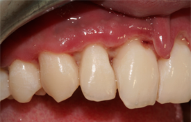 Figure 1: Generalized punched out papillae with pseudomembrane. Detached midline papillae. (photo courtesy of Dr. Eraldo Batista)
 Figure 2: Mild NUG case with erythematous marginal and interproximal gingival with slightly cratered papillae.
Symptoms
- Intense/excruciating pain
- Predisposing factors:
- Psychological stress and anxiety
- Smoking
- Pre-existing gingivitis and trauma
- Poor oral hygiene
- Deficient nutrition
- HIV-positive
- All the factors above lead to immunosuppression: depressed polymorphonuclear leukocytes, antibody response, and lymphocyte mitogenesis.
Investigation
- Thorough medical history, including nutrition and health habits
- Medical consult if immunosuppressive disease is suspected
- Dental history; pain (constant, intense onset)
- Extraoral examination; look for lymphadenopathy of the head and neck
- Intraoral examination; look for clinical features of NUG and presence of pasty saliva
Diagnosis
Based on the clinical examination, a diagnosis of NUG is determined.
Differential Diagnosis
- Primary herpetic gingivostomatitis
- Desquamative gingivitis
- Agranulocytosis
- Cyclic neutropenia
- Leukemia
- Ascorbic acid deficiency and gingivitis
Treatment
Common Initial Treatments
- Perform debridement under local anesthesia
- Remove pseudomembrane using cotton pellet dipped in chlorhexidine
- Provide patient with specific oral hygiene instructions to use a prescription antibacterial mouthwash: chlorhexidine 0.12% twice daily
- Control pain with analgesics: ibuprofen 400-600 mg 3 times daily
- Patient counselling should include instruction on proper nutrition, oral care, appropriate fluid intake, and smoking cessation
- Prescribe antibiotics if patient is immunocompromised (e.g., AIDS, leukemia, cyclic neutropenia) or in case of systemic involvement like fever, malaise and lymphadenopathy
- Follow up with a comprehensive periodontal evaluation after resolution of the acute condition
- For any signs of systemic involvement, the recommended antibiotics are:
- Amoxicillin, 250 mg 3 x daily for 7 days and/or
- Metronidazole, 250 mg 3 x daily for 7 days
- Assess treatment outcomes in 24 hours, then every other day until signs and symptoms are resolved and gingival health and function are restored (Figs. 3 and 4).
- Residual interdental soft tissue craters are more susceptible to further clinical attachment loss; evaluate possible surgical treatment of these areas.
- Sites that are nonresponsive to treatment may occur and may be characterized by recurrence and/or progressive destruction of the gingival and periodontal attachment.
- Reasons for nonresolution include the failure to remove the causes of irritation, incomplete debridement, inaccurate diagnosis, patient noncompliance, and/or underlying systemic conditions.
- Additional therapy and/or medical/dental consultation may also be indicated for nonresponding patients. These conditions may have a tendency to recur; therefore, frequent periodontal maintenance visits and meticulous oral hygiene are necessary.
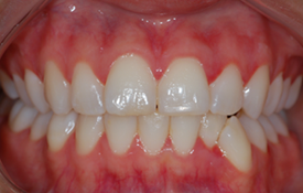 Figure 3: 24 h post treatment presenting reduction of erythematous margins and edema.
 Figure 4: Closer view 24 h after treatment.
Advice
- Untreated, the infection may lead to rapid destruction of the periodontium (necrotizing ulcerative periodontitis) and can even spread, as necrotizing stomatitis or noma, into neighbouring tissues in the cheeks, lips or the bones of the jaw.
- With treatment, even if clinical attachment loss is associated with NUG, resolution after treatment (periodic scaling, root planing and antimicrobial rinses), is quick and regeneration of the affected interdental soft tissues is possible.
THE AUTHORS
 |
Dr. Atout is an assistant professor in periodontology, division of periodontics, department of dental diagnostic and surgical sciences, faculty of dentistry, University of Manitoba, Winnipeg, Manitoba. |
 |
Dr. Todescan is an assistant professor in periodontology, division of periodontics, department of dental diagnostic and surgical sciences, faculty of dentistry, University of Manitoba, Winnipeg, Manitoba. |
Correspondence to: Dr. Reem Atout, Dental Diagnostic Surgical Sciences, D344 Dental Building, 790 Bannatyne Avenue, Winnipeg, MB, Canada, R3E 0W2. Email: reem.atout@ad.umanitoba.ca
The authors have no declared financial interests.
This article has been peer reviewed.
Suggested Resources
- Carranza N. Carranza’s Clinical Periodontology. 11th ed. St. Louis: Saunders, 2012. p.97-101,181.
- Corbet EF. Diagnosis of acute periodontal lesions. Periodontol 2000. 2004;34:204-16.
- Novak MJ. Necrotizing ulcerative periodontitis. Ann Periodontol. 1999;4(1):74-8.