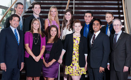
 Front row (l. to r.): Dave Hancin, vice-president and general manager, DENTSPLY Canada; Stephanie Smith, Dalhousie University; Camille Ruel, Laval University; Allison Nicholls, University of British Columbia; Jay Lalli, University of Saskatchewan; Dr. Robert Sutherland, CDA president. Back row (l. to r.): Kristopher Scott Currie, University of Saskatchewan; Matthew Morrison, McGill University; Patricia Brooks, Schulich School of Medicine and Dentistry; Janelle Hamilton, University of Montreal; Stephen Spano, University of Toronto; Matthew Kotyk, University of Manitoba.
The 2012 CDA/DENTSPLY Student Clinician Research Program took place in Saskatoon during the annual CDA Conference. This national clinical research competition invites dental students from the 10 accredited Canadian dental schools to present research table clinics in front of qualified judges.
Matthew Morrison of McGill University was named the winner of the 2012 program for his case-control study on the association between oral tori and local mechanical and systemic factors. His first prize is an expense-paid trip to 2012 American Dental Association (ADA) annual meeting in San Francisco, where he will present his research during the ADA’s scientific program.
“It was truly a humbling experience to speak with dental professionals regarding my research, and I look forward to presenting at this year’s ADA session,” says Dr. Morrison.
The runner-up in this year’s competition was Patricia Brooks of the Schulich School of Medicine and Dentistry at Western University. Her research examined cross-talk between signalling pathways in osteoblasts. “The conference was a remarkable opportunity to meet fellow dental student researchers from all across Canada. Having this experience, to establish relationships with like-minded students with a passion for research so early in each of our dental careers, will definitely help facilitate future collaborations,” she says. Ms. Brooks received a $1000 cash award from DENTSPLY for her second-place finish.
DENTSPLY has sponsored the Student Clinician Research Program in Canada for more than 40 years. The program’s purpose is “to stimulate ideas, to improve communication and most of all, to increase student involvement in the advancement of the dental profession.”
The student clinicians participating in the event are also honoured by the Canadian section of the Pierre Fauchard Academy (PFA). The students are presented with a scholarship from the PFA recognizing their special efforts in the advancement of dental education over and above their academic careers.
++++++
JCDA is pleased to publish condensed versions for the abstracts submitted for the CDA/DENTSPLY 2012 Student Clinician Research Program. To qualify, the study must fall under the “clinical application and techniques” or “basic science and research” categories. Students must identify the purpose of the study, provide background information, outline how the study was conducted and report on the results of the study and its possible significance. The student, selected by his or her own faculty, must be an undergraduate at the time of his or her selection. Nine dental schools participated in this year’s competition.
The abstracts are published in the language of submission.
++++++
1st Place
Oral tori are associated with local mechanical and systemic factors: a case-control study
By: Matthew Morrison, McGill University
Advisor: Dr. Faleh Tamimi
 Matthew Morrison
Despite the high prevalence of oral tori, an accepted mechanism for their development is not known. Understanding the processes involved in ectopic oral bone formation, as with oral tori, could assist in developing less invasive methods for alveolar ridge augmentation and natural enhancement of bone quality for dental implantation. Recent research has correlated mechanical stress, low-grade hyperactivity of the immune system, and treatment with hypertension medications with alterations in bone metabolism favouring bone apposition. Accordingly, we hypothesized that dental factors, medical conditions, and pharmacological agents known to stimulate bone formation would be associated with an increased risk for the presence of oral tori.
Using a case-control study design, we identified and adjudicated a sample of cases (with torus palatinus and/or torus mandibularis) and controls during a 1.5-year period. The medical records were abstracted and data on dental factors, temporomandibular dysfunction (TMD), medications, and medical conditions were recorded.
Our findings demonstrated indirect support for the role of bruxism in the development of oral tori. We also found a strong association between subjects with TMD and the presence of oral tori. Furthermore, a significant association was observed between treated hypertension and the presence of torus palatinus, as well as between subjects with a penicillin allergy (low-grade hyperactivity of the immune system) and the presence of torus mandibularis. Overall, our work identified relationships between ectopic oral bone formation and various patient characteristics involving local mechanical factors, the sympathetic nervous system, and the immune system.
2nd Place
Cross-talk between Wnt/β-catenin and P2X7 nucleotide receptor signalling pathways in osteoblasts
By: Patricia J. Brooks, Schulich School of Medicine and Dentistry at Western University
Advisor: Dr. Jeff Dixon
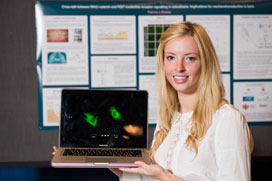 Patricia J. Brooks
The Wnt/ß-catenin and P2X7 nucleotide receptor signalling pathways are important for bone formation and mechanotransduction. Our objective was to investigate if cross-talk occurs between these pathways in osteoblasts.
MC3T3-E1 osteoblast-like cells were treated with Wnt ligand(Wnt3a), the P2X7 agonist BzATPor their controls, alone or in combination. At time points 0-24 h, samples were fixed, immunostained for ß-catenin and visualized by confocal microscopy. The localization of ß-catenin was quantified as an indicator of ß-catenin activation. To assess transcriptional activity directly, cells were transfected with a ß-catenin luciferase reporter plasmid and treated with Wnt3a or BzATP.
Wnt3a alone elicited a gradual increase of ß-catenin nuclear localization that peaked at 3 h and returned to baseline by 24 h. In contrast, BzATP caused transient nuclear localization only at 30 min. Notably, when cells were treated with Wnt3a+BzATP, nuclear localization of ß-catenin was more rapid and sustained compared to Wnt3a alone. Moreover, the intensity of nuclear localization was significantly higher in cultures treated with Wnt3a+BzATP, compared to Wnt3a alone. The reporter assay revealed that BzATP alone did not increase b-catenin transcriptional activity, whereas Wnt3a+BzATP elicited greater ß-catenin activity than Wnt3a alone, establishing that P2X7 activation enhances Wnt/ß-catenin function in osteoblasts.We have shown for the first time in any system that P2X7 activation prolongs and potentiates ß-catenin activity elicited by Wnt signalling, thereby establishing existence of cross-talk between these two important pathways.Thiscross-talk may enhance the anabolic response of bone to mechanical stimulation asoccurs during distraction osteogenesis and orthodontic tooth movement.
A retrospective study examining factors influencing patient compliance in a periodontal maintenance program and its influence on tooth loss and clinical attachment level
By: Janelle Hamilton, University of Montreal
Advisors: Drs. Robert Durand and René Voyer
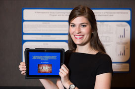 Janelle Hamilton
The aims of this retrospective study were to determine factors influencing patient compliance and whether patient compliance in a periodontal maintenance therapy (PMT) program influences tooth loss and clinical attachment level.
Data from 759 patients who were treated by a periodontist between 1969 and 2003 were reviewed. One hundred and twenty patients diagnosed with chronic periodontitis were included in this study. Subjects were at least 35 years old, underwent active periodontal therapy and were maintained for a minimum of 5 years. Patient-level risk factors and clinical parameters, including tooth loss throughout periodontal maintenance therapy, were collected. Data were analyzed to detect differences between complete and erratic compliers using 2 different compliance definitions (1 and 2) previously described in the literature.
There were no significant differences in patient-specific risk factors for periodontitis between complete and erratic compliers 2, except for bruxism (p = 0.005). There were no significant differences in the number of teeth lost in complete vs. erratic compliers 2 ( p= 0.65). Complete compliers 2 had significant improvements in mean pocket depths compared to erratic compliers 2 (p = 0.007). However, no differences in mean clinical attachment level change were found between the two groups (p = 0.16).
There were no differences in tooth loss between complete and erratic compliers enrolled in a PMT program. Although complete compliers exhibited higher mean pocket depth reduction, no significant differences were found for clinical attachment level. This study highlights the need for more prospective studies of PMT measuring patient compliance and clinical outcomes.
An investigation into bisphenol A (BPA) leaching from orthodontic materials
By: Matthew Kotyk, University of Manitoba
Advisor: Dr. William Wiltshire
 Matthew Kotyk
In clinical dentistry, numerous types of dental materials are in direct contact with oral tissues and oral fluids. The leaching of components from these dental materials could be a clinical concern. Within resin composite and plastic materials, a leached component of particular interest is bisphenol A (BPA) due to its cytotoxic and estrogenic properties. This study investigated BPA leaching from orthodontic materials including, but not limited to, clear aligners when subjected to the most extreme conditions experienced intra-orally, including thermal shock and abrasion.
As-received orthodontic materials used in routine orthodontic treatment were obtained from the manufacturers, with one exception. All materials were subjected to vigorous thermal and physical conditions to identify if those conditions would be responsible for stimulating BPA release. The standard leaching conditions consisted of initial surface abrasion followed by immersion in a medium of artificial saliva, followed by thermal shock treatment involving temperature cycling from hot to cold with shaking for 5 minutes at each temperature and repeated for a total of 10 cycles. The samples were then shaken at normal oral temperature and aliquots removed at regular time intervals. All aliquots were analyzed for BPA.
The conditions to which the orthodontic materials were subjected in this study represent the extremes of those encountered intra-orally. For some materials, no BPA release was detected and the concerns with regards to those materials are alleviated. Conversely, the discovery of BPA leaching from some materials will necessitate a review of their use.
The effect of back fitness on changes in muscle activation and fatigue after performing a dental procedure
By: Jay Lalli and Kristopher Scott Currie, University of Saskatchewan
Advisor: Dr. Carol Lynn Nagle
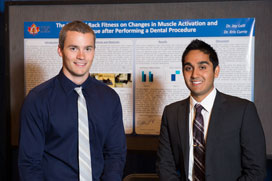 Kristopher Scott Currie and Jay Lalli
Dental professionals are prone to back muscle injuries due to the inherent occupational stresses within the profession. Our main objective was to determine whether dental students with low back fitness experience greater muscle fatigue and strain compared to students with high back fitness, while the secondary objective was to gain insight into the factors that may play a role.
Twenty-nine dental students consented to participate in this study. Students completed the Waterloo Handedness Questionnaire, Physical Activity Readiness Questionnaire and the Canadian Society for Exercise Physiology back fitness test, which established their back fitness score. Participants were attached to an electromyogram (EMG) and baseline readings of the erector spinae were established. The participants performed a 15-minute tooth preparation, with recordings before and after the procedure, as well as a maximal voluntary contraction reading. Muscle activation and median EMG frequency were compared between participants with high and low scores.
Participants with low back fitness scores displayed increased EMG activity before and after the procedure when compared to those with high back fitness, indicating overall greater strain on the erector spinae. The lower back fitness group experienced a decreasing median frequency, indicating increased muscle fatigue. Participants with low back fitness scores experience greater muscle strain and fatigue of the erector spinae during dental procedures. Dentists should take an active role in preventing back strain and fatigue, such as exercises that improve muscular endurance and reduce waist girth, to ensure healthier careers in dentistry.
Kinetic characterization of a novel class of anticollagenase catK inhibitors
By: Allison Nicholls, University of British Columbia
Advisor: Dr. Ravindra Shah
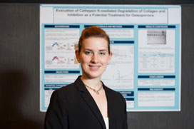 Allison Nicholls
Osteoporosis is a bone degenerative disease caused by excessive osteoclast activity. Cathepsin K is a cysteine protease that is highly expressed in osteoclasts. This enzyme forms a complex with chondroitin 4-sulphate (C4-S) creating collagenase activity. The objective of the research was to determine the kinetic parameters of collagen hydrolysis by cathepsin K and to evaluate the potency of previously identified cathepsin K/C4-S complex formation inhibitors.
Km and kcat values were determined for the degradation of type-I collagen by the cathepsin K/C4-S complex. Michalis-Menten kinetics and a Lineweaver-Burk analysis were used to evaluate the data. Structurally-related putative cathepsin K/C4-S complex formation inhibitors were evaluated for their ability to prevent cathepsin K/C4-S-mediated degradation of collagen in a non-active site inhibitory mechanism. An IC50 was determined for each effective compound, and the accessibility of the active site of cathepsin K was determined using a synthetic fluorogenic peptide substrate, Z-FR-MCA.
The kinetic values for cathepsin K/C4-S-mediated degradation of collagen are as follows: Km = 1.6 mM, kcat = 6 h-1 and kcat/Km = 1.062 M-1s-1. Six out of the 8 putative cathepsin K inhibitors were able to prevent the degradation of collagen: aurintricarboxylic acid (IC50 = 9.6 μM); ellipticine (IC50 = ~100 μM); epigallocatechin gallate (IC50 = 52 μM); raloxifene (IC50 = 85 μM); suramin (IC50 = 6.7 μM); and tamoxifen (IC50 = ~150 μM). All but suramin appeared to be non-active site inhibitors. Interestingly, tamoxifen and raloxifene have been clinically demonstrated to inhibit bone resorption via an estrogen-related pathway.
L’effet des diètes molles sur la guérison tissulaire gingivale après une chirurgie buccale : une étude in vitro
By : Camille Ruel, Laval University
Advisor: Dr. Lise Payant
 Camille Ruel
L’objectif général était d’analyser l’impact positif ou négatif de certains aliments présents dans les diètes molles sur le processus de guérison tissulaire gingivale et d’identifier un moyen pour favoriser la cicatrisation d’une lésion buccale à la suite d’une chirurgie, sans pour autant affecter les conditions de vie normale du patient. Les résultats obtenus permettront donc d’améliorer les recommandations formulées aux patients à la suite d’une intervention chirurgicale.
Trois aliments ont été testés sur des cellules gingivales humaines (épithéliales et fibroblastes) : un jus d’orange, un substitut de repas ainsi qu’un yogourt à boire. Trois types d’expérimentation ont été réalisés afin de pouvoir analyser les effets des produits sur la migration, la prolifération et la différenciation cellulaire, et ce, à différents pH. Soit la première série d’expériences aux pH normaux des différentes solutions et la seconde série à des pH normalisés à 6,8.
Les résultats obtenus montrent en général que seul le substitut de repas ne semble pas nuire à ces 2 phénomènes lorsque les produits ne sont pas modifiés. Il a été également déterminé que le pH des aliments influence grandement leur effet nocif sur les cellules gingivales. Donc, une première recommandation serait de ne pas consommer d’aliments acides lors de la diète molle prescrite après une chirurgie buccale. Il ne va sans dire que plusieurs autres tests devront être réalisés avant de vraiment pouvoir transposer ces résultats in vivo et obtenir de cette recherche un usage clinique pour les professionnels du domaine dentaire.
Current choices for all ceramic restorations: evidence-based review with clinical guidelines for material selection
By: Stephanie Smith, Dalhousie University
Advisor: Dr. Mark Vallee
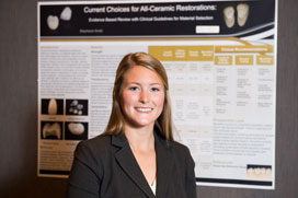 Stephanie Smith
Currently, there is an expansive selection of all ceramic materials; however, none encompass the ideal properties of strength and esthetic appropriate for all clinical situations. Therefore, the purpose of this research project is to provide relevant comparisons between current glass and zirconia based all-ceramics, as well as provide clinical recommendations for material use.
A literature review was completed with the PubMed database including the search terms “ceramics,” “dental porcelain,” “leucite,” “lithium disilicate,” and “zirconia.”
Searches were limited to clinical trials, meta-analyses, randomized control trials and papers written in English.
Research revealed that zirconia based all-ceramics exhibit high flexural strength and fracture toughness, but are limited by their translucent and therefore esthetic properties. Alternatively, glass-based ceramics exhibit high translucency but have weaker physical properties of strength and fracture toughness. These variations in material properties influence which all-ceramic is appropriate for differing clinical applications. Due to their increased esthetic properties but weaker clinical strength, glass-based ceramics have been recommended for in lays/on lays, anterior and posterior single crowns, and anterior fixed partial dentures (FPDs). Zirconia coping restorations combine superior mechanical properties with esthetic overlying porcelain, therefore these materials are recommended for anterior and posterior single crowns and FPDs, as well as locations to conceal an underlying restoration. Finally, single layered monolithic zirconia is recommended for posterior single crowns and FPDs, areas of parafunction, limited interarch space and locations to conceal underlying restorations.
Role of discoidin domain receptor 1 in cyclosporine A-induced gingival overgrowth
By: Stephen Spano, University of Toronto
Advisor: Dr. Christopher McCulloch
 Stephen Spano
Gingival overgrowth is an adverse outcome of cyclosporine A (CsA) treatment, involving disruption of collagen remodeling. Fibroblast receptors that mediate remodelling and that are dysregulated by CsA are not defined. We hypothesized that discoidin domain receptor 1 (DDR1) expressed by fibroblast is important for collagen remodeling and that DDR1 is affected by CsA, thereby disrupting collagen homeostatis.
Our objectives were to (1) determine whether DDR1 mediates collagen remodeling by fibroblasts; (2) assess the effets of CsA on DDR1-mediated collagen remodeling.
Mouse 3T3 fibroblast cells expressing DDR1 or null for DDR1 were treated with vehicle (dimethyl sulfoxide-DMSO) or CsA (1 µM in DMSO). Cells were inoculated into floating or attached collagen gels to study the role of DDR1 in fibrillar reorganization or cell-mediated traction, respectively. Cell migration was analyzed with in vitro scratch wound assays.
DDR1-expressing cells contracted floating gels more rapidly than DDR1 null cells (mean slope±S.E.M.: DDR1=75.4±12.2 µm/hr; DDR1 null cells=18.9±4.8 µm/hr; p < 0.001). DDR1-expressing cells treated with CsA exhibited a 2.2-fold reduction of gel contraction (34.4±2.3 µm/hr; p < 0.001). CsA has no effect on DDR1 null cells (p > 0.0.5). In attached collagen gel concentration assays, DDR1-expressing cells contracted gels more rapidly than DDR1 null cells (877±63 µm/hr vs. 247±87 µm/hr; p < 0.001). CsA did not affect attached gel concentration. DDR1 expression and CsA did not affect cell migration in scratch wound assays (p > 0.0.5).
DDR1 is important for collagen remodeling by fibrillar reorganization and by contraction in vitro, but CsA only affects fibrillar reorganization. Accordingly, CsA perturbation of DDR1-mediated fibrillar reorganization may contribute to gingival overgrowth.
Photos courtesy of Teckles Photos Inc.
